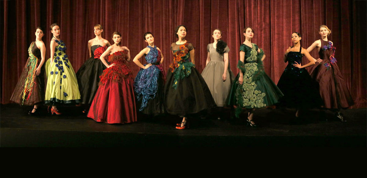
Models of the Fashioning Cancer Collection. Photo credit: Tim Matheson
FASHIONING CANCER:
THE CORRELATION BETWEEN
DESTRUCTION AND BEAUTY
March 25, 2014 | 12-1pm
Free Public Presentation
Graduate Research Seminar Series:
Frederic Wood Theatre, UBC
If you are a woman who has battled cancer we would very much appreciate knowing your response to the gowns, negative or positive. Please send your comments to Associate Professor Jacqueline Fikins at jacqueline.firkins @ ubc.ca and indicate if you would consent to have your comments included in her research paper when goes to print at the end of March, 2014.
Fashioning Cancer: The Correlation between Destruction and Beauty is a “dramatic” new interdepartmental research project at UBC made possible through a Research Mentorship Grant from the Peter Wall Institute For Advanced Studies
Watch the Canadian Press Video »
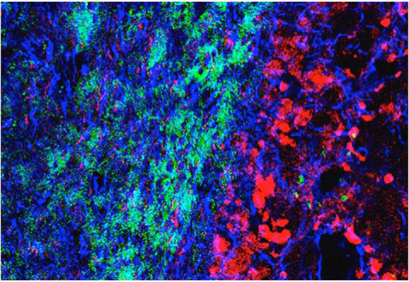
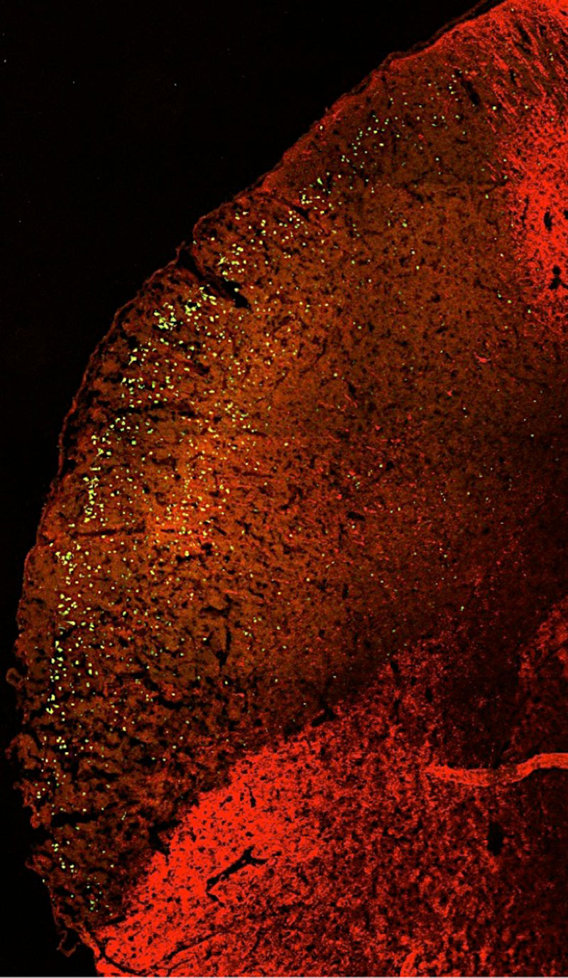
Normal brain area on the left (blue and green) encountering invading cancer cells (glioma; red). Studying how cancer cells interact with normal cells is key to understanding ways to prevent invasion and metastasis of cancer cells.
Photo Credit: Wun Chey Sin, Ph.D., Christian Naus, Ph.D.
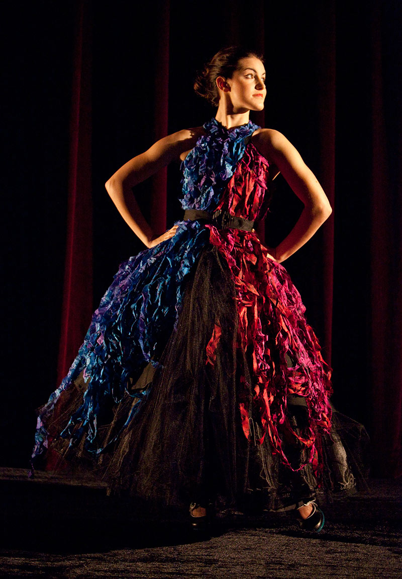
(Model) Eva Tavares | UBC Opera student. Black faille halter w/ blue/pink silk shredded trim & tulle underskirt black silk faille with shredded vintage silk saris, tulle underskirt over sparkle organza.
Photo credit: Tim Matheson
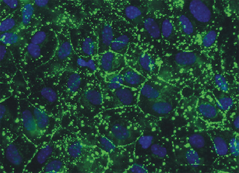
Astrocytes growing in a culture dish. Blue dye indicated the nuclei (DNA); green color shows the location of gap junction channels which link the cells in a metabolic community. These channels are important in controlling the normal growth of cells; their disruption can promote cancer development.
Photo Credit: Hoa Le, Ph.D., Christian Naus, Ph.D.
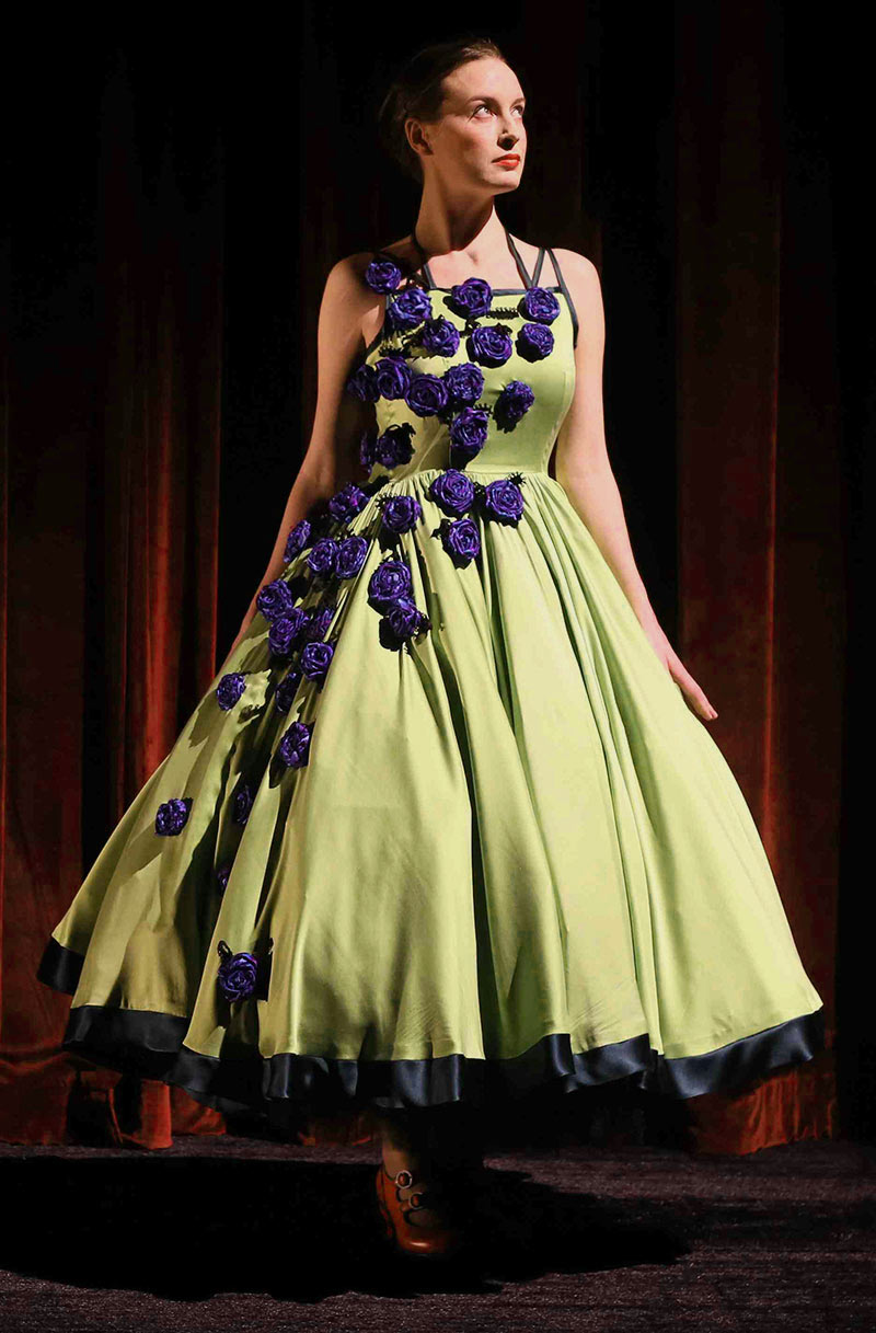
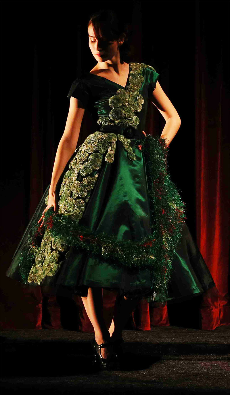
(Model) Bronwyn Malloy | UBC Alumna, McGill MA English student, and UBC PhD English student (Fall 2014). Green silk charmeuse w/blue rosettes & dark green hem/edging.
Photo credit: Tim Matheson
Brain tissue showing an area injury (faded red) which is filled with dying neurons (stained green). As cancers progress the normal cells die as part of disease progression.
Photo Credit: Moises Freitas-Andrade, Ph.D., Christian Naus, Ph.D.
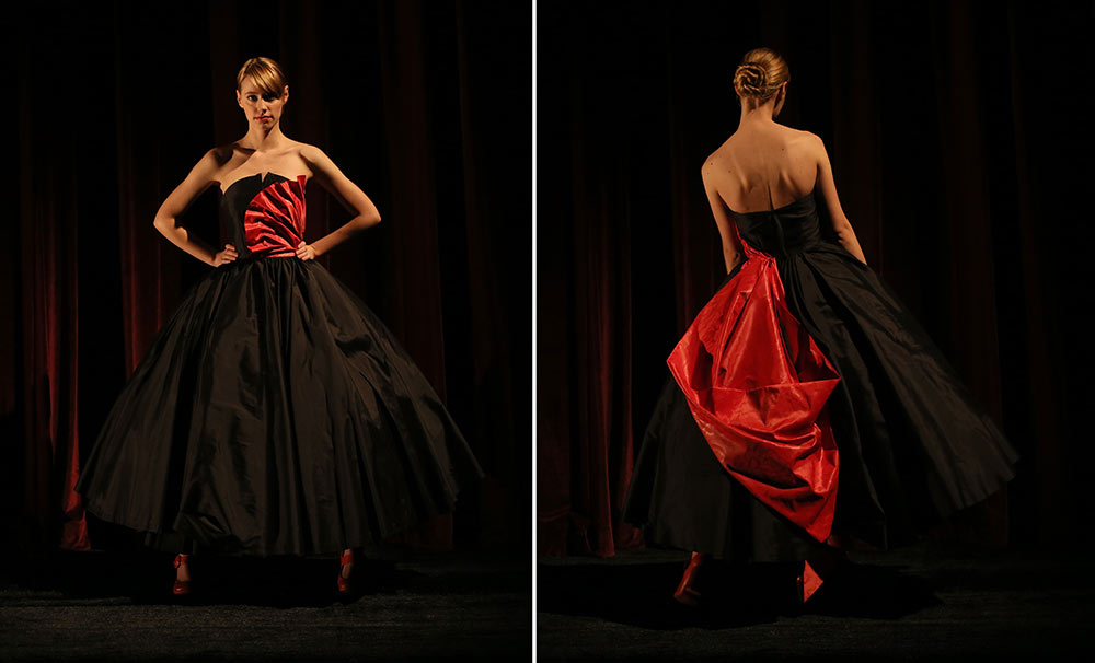
(Model) Rebecca Burks | UBC BFA Theatre Production & Design student. Black silk taffeta, red watered silk taffeta bow.
Photo credit: Tim Matheson
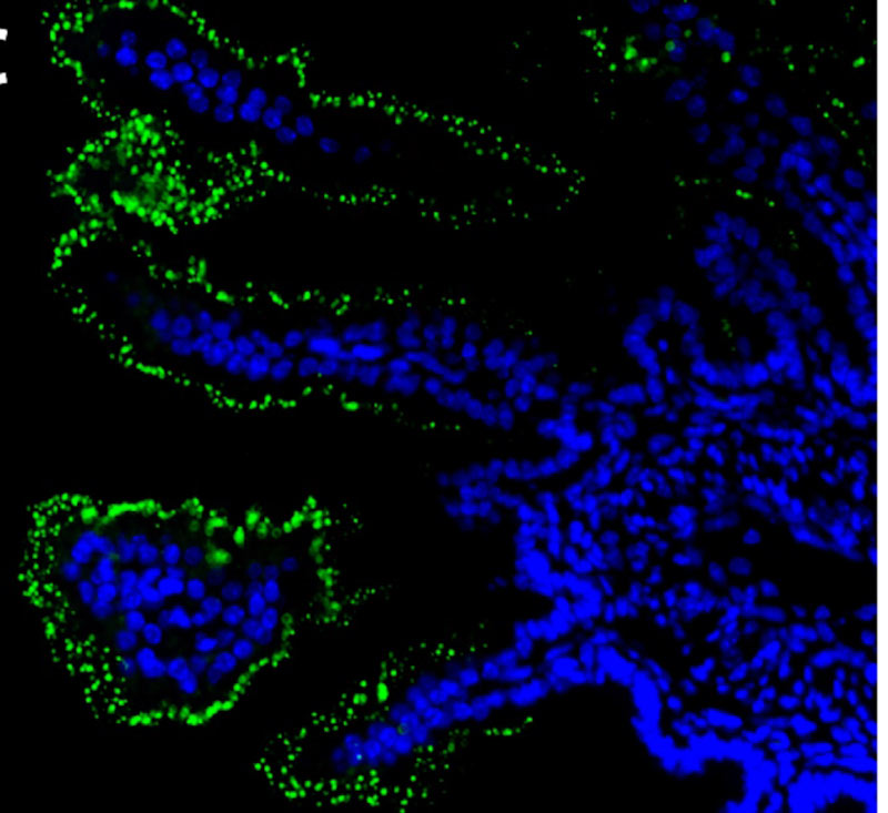
Small instestine – blue color shows the cell nuclei (DNA) and green color is localized to membrane channels called pannexins. Membrane channels like these contribute to the microenvironment that can either enhance or inhibit cancer progression.
Photo Credit: Stephen Bond, Ph.D., Christian Naus, Ph.D.
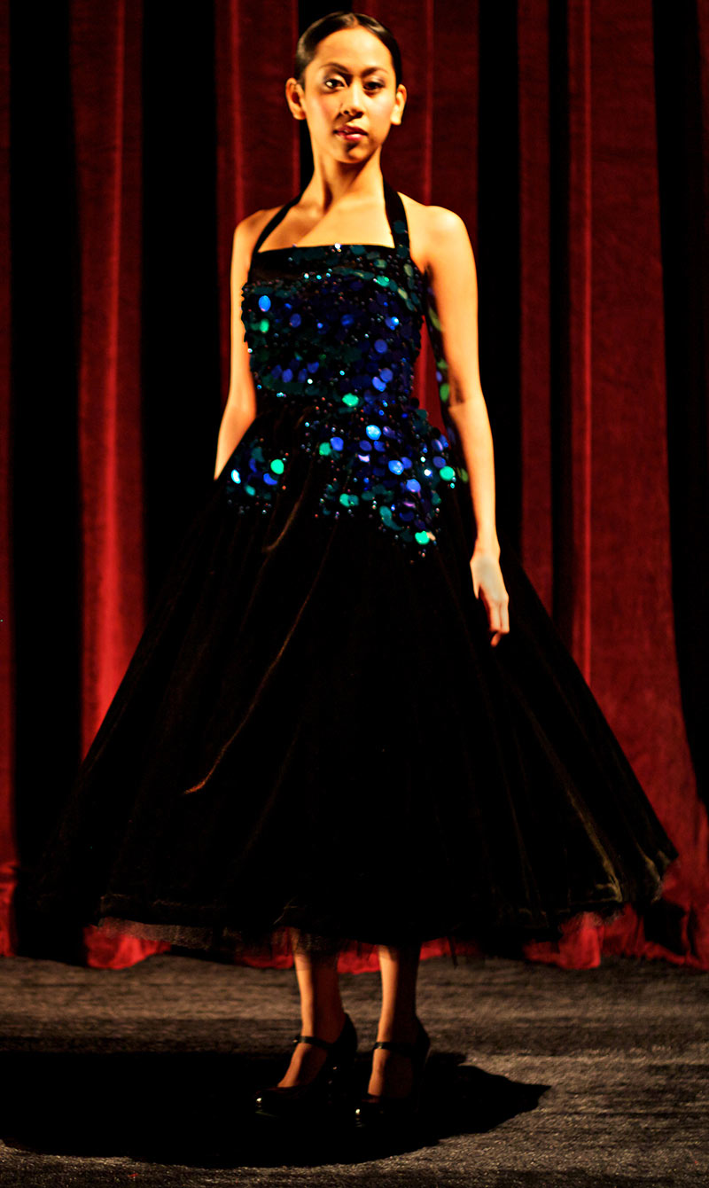
(Model) Sarah Roa | UBC BFA Acting student. Black velet halter with sequins & beads.
Photo credit: Tim Matheson
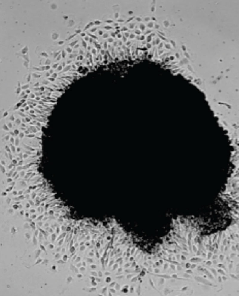
Brain tumor (glioma) growing in a culture dish. Individual cells are actively moving away from the tumor mass (black area) recapitulating what they do when invading the brain.
Photo Credit: Wun Chey Sin, Ph.D., Christian Naus, Ph.D.

(Model) Helena Fisher-Welsh | UBC BFA Acting student. Grey organza w/ black trim.
Photo credit: Tim Matheson
NOTE: Not all the slides show cancer – only #4 & 8 in this selection (above).
The following slides are examples of cellular systems that UBC cancer researchers are studying because they are subject to having their modes of activity hijacked by cancer to spread itself. All photos of cells were taken by UBC researchers in their work (see photo credits).

During brain development, nerve cells (neurons; green) migrate along supporting cells (radial glia; red) to specific coordinates in time and space in order to develop a network on interconnections with other neurons. Similar processes are used by cancer cells when they invade normal tissue and progress to metastatic disease.
Photo Credit: Christian Naus, Ph.D., Cima Cina, Ph.D.
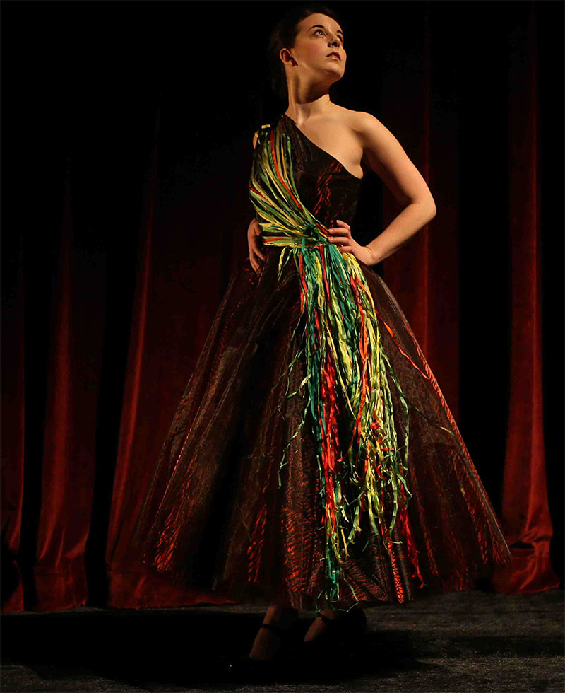
(Model) Simone McIntosh | UBC Opera student. Zebra print organza with single shoulder & silk ribbons.
Photo credit: Tim Matheson
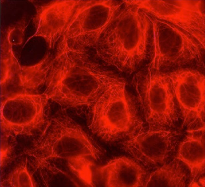
Astrocytes cells from the brain ensure that neurons remain healthy. can become cancerous and result in gliomas. These cells are stained to show their filamentous “cytoskeleton” which determines their shape as well as impacting their ability to move. In cancer cells, the more they move, the more aggressive the disease.
Photo Credit: John Bechberger, M.Sc.; Christian Naus, Ph.D.

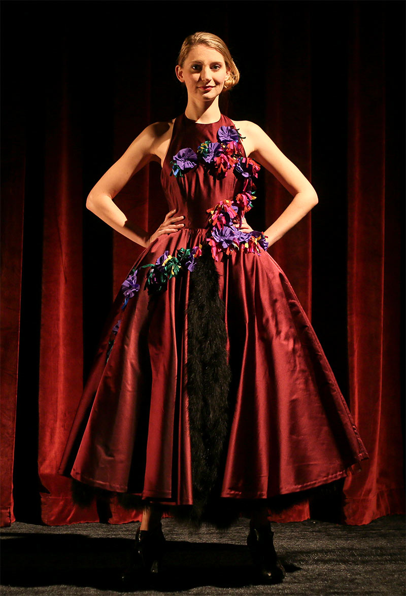
(Model) Chelsi Walsh | UBC MFA Opera student. Red taffeta strapless full length with yarn & eyelash top.
Photo credit: Tim Matheson

Astrocytes from the brain growing in a culture dish. Green color indicates the cytoskeleton of these cells; red color shows specific membrance channels (gap junctions); blue color indicates the cell nuclei (DNA). The ability to grow cells in a dish has contributed to our understanding of the changes theses cells undergo when they become cancerous.
Photo Credit: John Bechberger, M.Sc., Christian Naus, Ph.D.
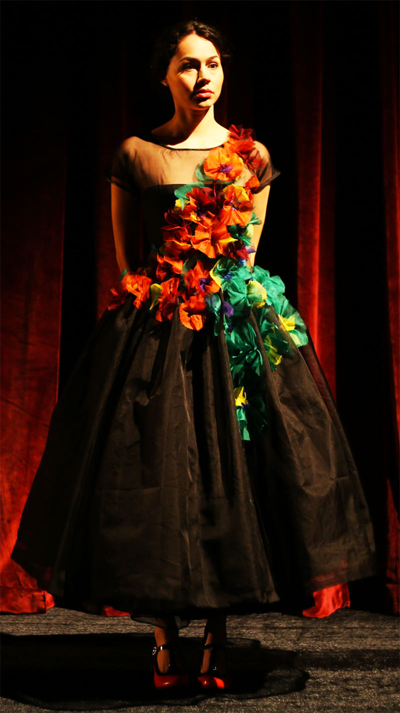
(Model) Mercedes de la Zerta | BFA Acting student. Black organza cap sleeve w/ sheer top & multicolour organza diagonal trim.
Photo credit: Tim Matheson
Brain cancer cells showing interaction of two proteins – gap junction channels (red) and a cell growth factor (green). Yellow indicates areas of the cell where both proteins interact. These types of interactions between proteins are critical in processes which can transform normal cells into cancer.
Photo Credit: Christian Naus, Ph.D.
(Model) Chanel McCartney | UBC MFA Costume Design student. Green taffeta short sleeve with speckled applique diagonal trim & eyelash scarf.
Photo credit: Tim Matheson

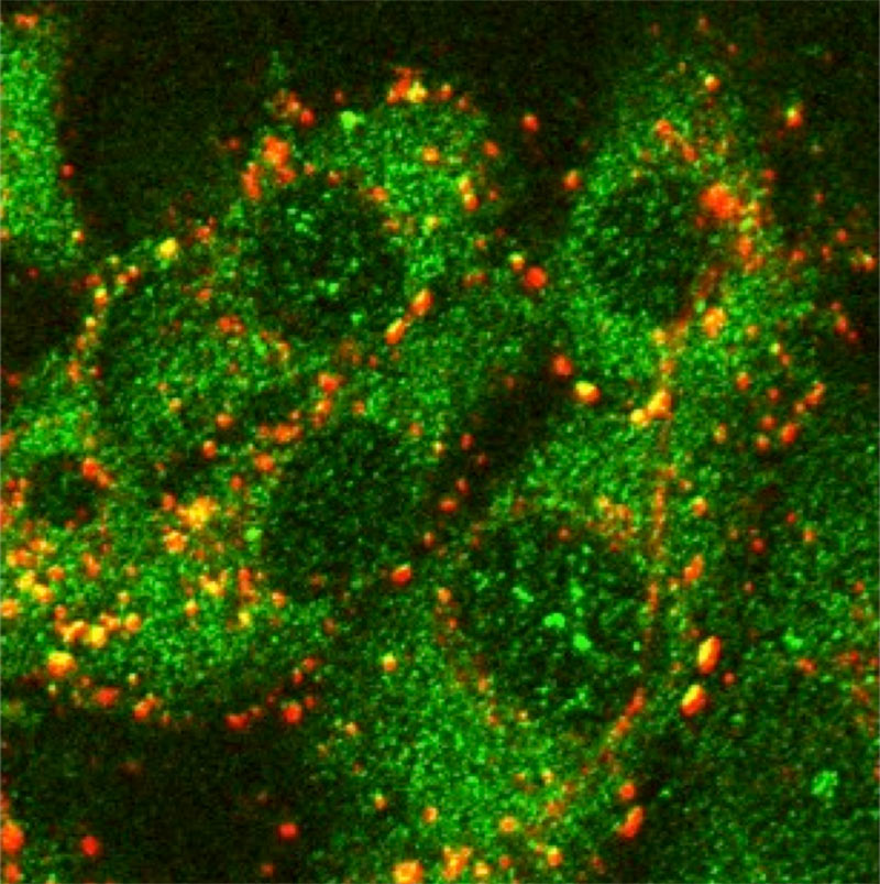
Brain cancer cells showing interaction of two proteins – gap junction channels (green) and a cell growth factor (red). Yellow indicates areas of the cell where both proteins interact. These types of interactions between proteins are critical in processes which can transform normal cells into cancer.
Photo Credit: Christian Naus, Ph.D., Christine Fu.
(Model) Katherine McLaughlin | UBC BFA Acting student. Purple satin halter with feather underskirt & diagonal purple rosettes with shreded bias streamers.
Photo credit: Tim Matheson